
More Information
Submitted: March 28, 2023 | Approved: April 13, 2023 | Published: April 14, 2023
How to cite this article: Ikejiri LLAA, Álamo L, Galli MZ, Bombonatti JFS, de Amoêdo Campos Velo MM, et al. Effectiveness, longevity, and color stability of in-office bleaching (6% H2O2 gel/Violet LED) and diastema closure with direct composite: 3-year follow-up. J Clin Adv Dent. 2023; 7: 001-006.
DOI: 10.29328/journal.jcad.1001033
Copyright License: © 2023 Ikejiri LLAA, et al. This is an open access article distributed under the Creative Commons Attribution License, which permits unrestricted use, distribution, and reproduction in any medium, provided the original work is properly cited.
Keywords: Bleaching agents; Composite resins; Diastema; Light source; Tooth bleaching

Effectiveness, longevity, and color stability of in-office bleaching (6% H2O2 gel/Violet LED) and diastema closure with direct composite: 3-year follow-up
Larissa Luri Almeida Amorim Ikejiri1 , Larissa Álamo1, Mateus Zamora Galli2, Juliana Fraga Soares Bombonatti1*, Marilia Mattar de Amoêdo Campos Velo1 and Rafael Francisco Lia Mondelli1
1Bauru School of Dentistry, Univesity of São Paulo, Department of Dentistry, Endodontics and Dental Materials, Bauru, Brazil
2Bauru School of Dentistry, Univesity of São Paulo, Department of Prosthodontics and Periodontology, Bauru, Brazil
*Address for Correspondence: Juliana Fraga Soares Bombonatti, Professor, Bauru School of Dentistry, Univesity of São Paulo, Department of Dentistry, Endodontics and Dental Materials, Bauru, Brazil, Email: [email protected]
To reduce bleaching side effects, the use of low concentrations of Hydrogen Peroxide (HP) agents associated with hybrid light (violet LED/Diode Laser) has gained interest.
Case report: The aim of this report is to describe a case of a 16-year-old patient that presented a complaint related to the color of his teeth and a maxillary midline diastema. In-office bleaching with 6% HP associated with hybrid light (violet LED/Diode Laser) was performed. The bleaching gel was applied once on the teeth and light-activated for 1 minute (15 times) followed by 1min intervals (15 times) with a total bleaching time of 30 minutes. After the bleaching procedure, the teeth were polished and the desensitizer was applied for 4 minutes. Two bleaching sessions were performed at a 1-week interval. The diastema was closed with direct resin composite restorations without any tooth preparation. The conventional 3-step bonding agent was used and A1 dentin shade and B1 enamel shade were used followed by polishing discs. At 3-year recall, discoloration and fractures were not found on the the teeth or restorations and patient was completely satisfied.
Conclusion: the conservative and safe option of bleaching with a low-concentrated HP gel associated with violet LED light is an interesting option for young patients and presents longevity over time.
The demand for aesthetic procedures in Dentistry is increasing since white and well-aligned teeth play an important role in a better overall facial appearance [1]. In this context, dental bleaching consists of a popular, effective and conservative treatment [2,3]. Hydrogen Peroxide (HP) is the most commonly used bleaching agent and is manufactured in concentrations ranging from 3% to 40% [4,5]. There are several protocols available for in-office bleaching: applying only peroxide bleaching gels; bleaching using only violet LED light or bleaching using HP gel associated with a light source [6,7]. The technique that uses only the bleaching gel is the most widely used because of the satisfactory results and no need of buying de light source equipment, which results in less treatment cost [8]. In this modality, the professional usually applies highly concentrated peroxide bleaching gel for a time ranging from 30 to 60 minutes [2,9] but is necessary two appointments at the least to obtain the dental bleaching.
Although high concentrations of HP in the whitening gel result in a faster bleaching outcome [4,10], the risk for postoperative sensitivity increases with concentration, once dental sensitivity is related to the diffusion of free radicals through enamel and dentin [3,11]. In addition, higher peroxide concentrations have been related to greater enamel surface alterations and cell damage [12,13].
For these reasons, new products and technologies have been developed for in-office dental bleaching. Recent protocols using low-concentrated peroxide bleaching gels have gained interest [14,15]. According to the Guide of the European Community [16], only bleaching products containing concentrations of >0.1 to 6% of HP present or released are considered safe for the patients. Keeping this context in mind, in order to maintain the whitening effect of highly concentrated bleaching gels and increase the safety of bleaching treatment, manufacturers proposed the incorporation of nanoparticles of titanium dioxide doped with nitrogen (TiO_N) photo-catalyst into lower concentrations of HP (3.5% – 6%) [17,18]. Researchers [19,20] demonstrated the effectiveness of 6% HP with nitrogen-doped titanium dioxide light-activated bleaching agent in clinical studies, with no difference in subjective color evaluations between 6% and 35% HP gels. The catalytic activity occurs when the TiO_N nanoparticles are exposed to wavelengths < 535 nm [20]; enhancing the generation of reactive oxygen species and improving the efficacy of bleaching gel [19,21]. The incorporation of catalytic nanoparticles allows 6%HP to still be effective with a reduced risk of sensitivity [20,22].
When considering a young patient, the use of a low-concentrated hydrogen peroxide agent seems to be an interesting option. The aim of this study is to illustrate a case report of effectiveness, longevity and color stability after 3 years of in-office bleaching with 6% HP gel doped with TiO_N, photocatalyzed with hybrid light (Violet LED/Diode laser) and diastema closure with direct resin composite in a 16-year-old patient.
A 16-year-old male patient was referred to the dental clinic of Bauru School of Dentistry (Bauru, SP, Brazil) with the aim of bleaching teeth and closing the midline diastema (Figure 1).
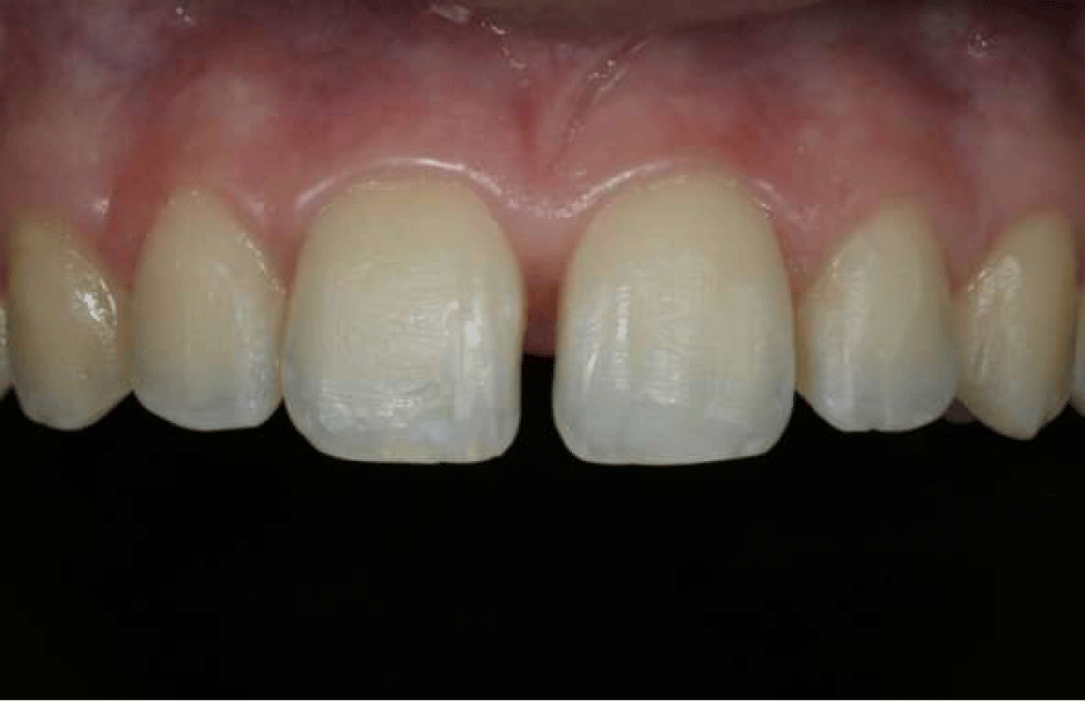
Figure 1: Initial aspect of the teeth.
The intraoral examination revealed that the diastema was due to the high labial frenal attachment. In addition to the labial frenectomy surgery, different treatment options were discussed to close the diastema. As the patient was only 16-year-old and had never been submitted to a bleaching procedure before, it was chosen the protocol of in-office bleaching with 6% HP gel doped with TiO_N (Nano white 6%, DMC Equipamentos Ltda, São Carlos, SP, Brazil) associated with hybrid light (violet LED/Diode Laser) (Whitening Lase Premium, DMC Equipamentos Ltda., São Carlos, SP, Brazil) before the restorative procedure of diastema closure. The choice of direct composite restoration technique was because of the need for the preparation of intact tooth surfaces.
The initial teeth shade A2 was determined using a classic shade guide (VITA, Bad Sackingen, Germany) (Figure 2).
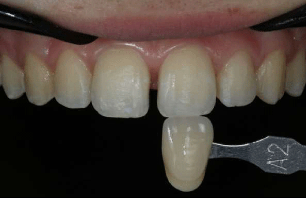
Figure 2: Initial color evaluation using VITA Classical Shade guide (A2).
Before the bleaching gel application, a prophylaxis procedure was done with pumice and the soft tissues were protected with a polymeric barrier (DMC Equipamentos Ltda., São Carlos, SP, Brazil). The bleaching protocol using 6% HP gel (Nano White, DMC Equipamentos Ltda., São Carlos, SP, Brazil) photocatalized with a hybrid light (violet LED/Diode Laser) (Whitening Lase Premium, DMC Equipamentos Ltda., São Carlos, SP, Brazil) was performed following the manufacturer’s instruction.
The bleaching gel was applied only one time onto the buccal surfaces of the isolated teeth and light-activated for 1 minute followed by 1min intervals without light until the completion of 15 light activations and 15 intervals with a total bleaching time of 30 minutes (Figure 3).
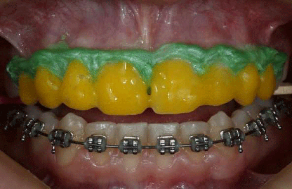
Figure 3: The 6% hydrogen peroxide gel on the tooth surface.
After the bleaching procedure, the teeth were then polished for the 20s with a wet felt disc impregnated for polishing (DMC Equipamentos Ltda., São Carlos, SP, Brazil) followed by the application of a desensitizer (Nano White, DMC Equipamentos Ltda., São Carlos, SP, Brazil) for 4 min. Two bleaching sessions were made at 1-week intervals without any tooth sensitivity. After 1 week of the second session, the patient was satisfied with the final color A1 (Figure 4), and the labial frenectomy surgery was made (Figure 5).
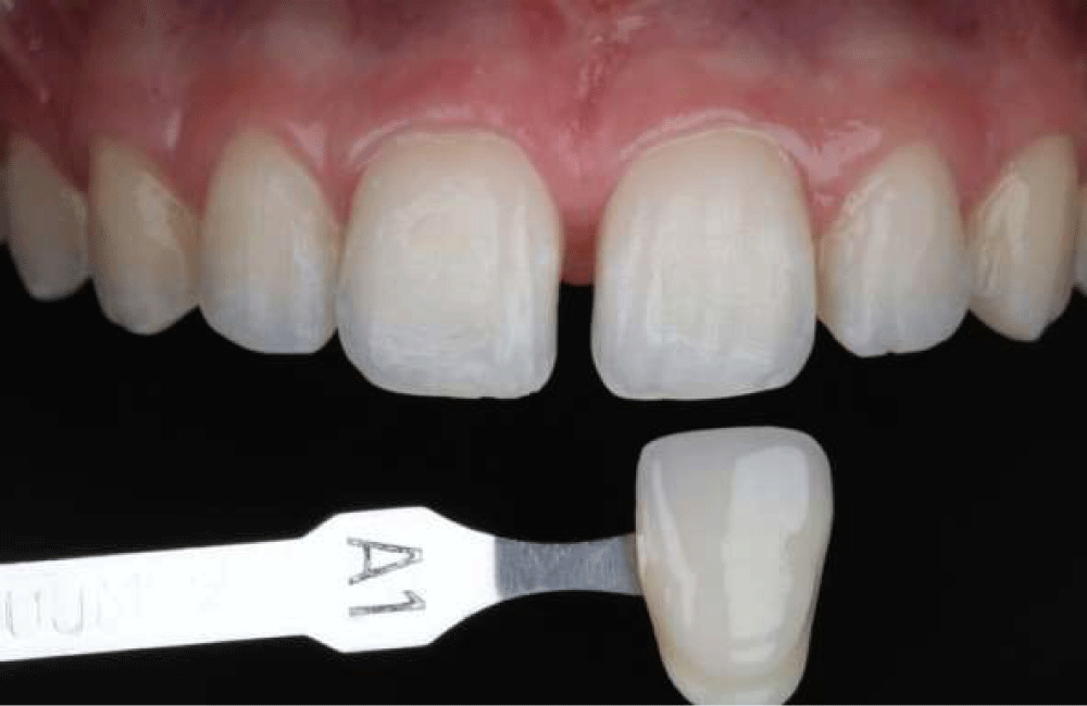
Figure 4: Final color evaluation using VITA Classical Shade guide (A1).
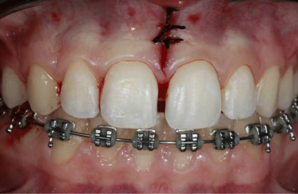
Figure 5: Immediate postoperative view after labial frenectomy surgery.
The diastema closure with direct resin composite restoration was performed 15 days after labial frenectomy. A resin shade test was done on the buccal surface of the maxillary incisor and the resins chosen for the restorative procedure were A1 for the dentin layer and B1 for the enamel layer (Vittra APS, FGM, Joinville, SC, Brazil). Then, all maxillary incisors were isolated with a rubber dam (Figure 6).
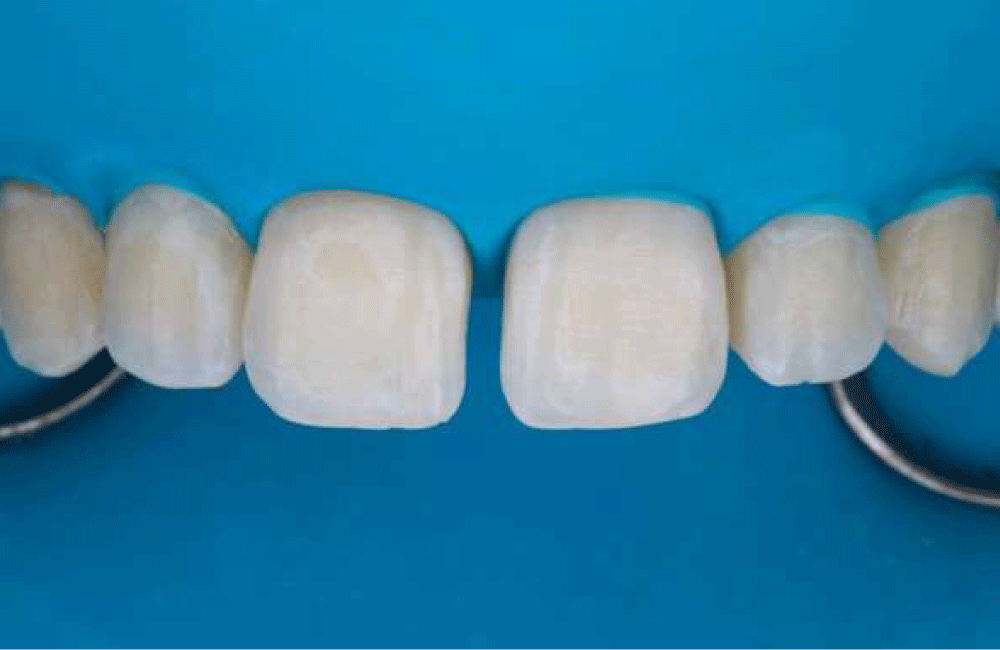
Figure 6: Maxillary incisors isolated with a rubber dam.
There was no teeth preparation before the restorative procedure. A 37% phosphoric acid (Condac, FGM, Joinville, SC, Brazil) was applied for 30 seconds on the mesial surface of the first teeth to be restored, while the adjacent incisor was covered with a Teflon band. After it was rinsed and dried for the same time, the bond of the conventional 3-step bonding agent (Scotch Bond Multi-Purpose Plus, 3M ESPE, St. Paul, MN, USA) was applied and polymerized for 30 seconds (Figure 7). After the first teeth (11) were restored (Figure 8), the same steps for hybridization were performed to restore the other incisor (21).
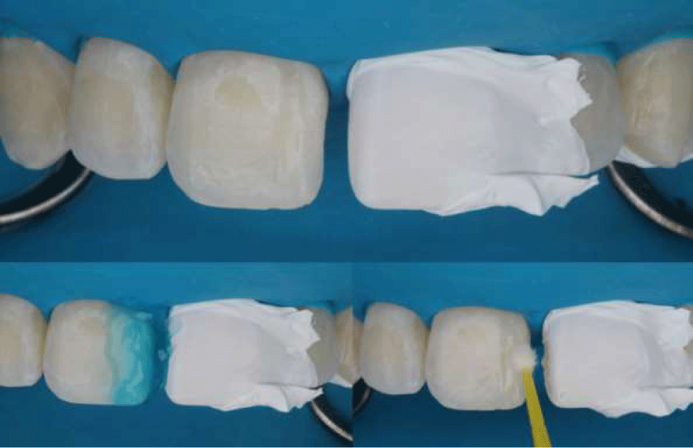
Figure 7: Hybridization of the enamel.
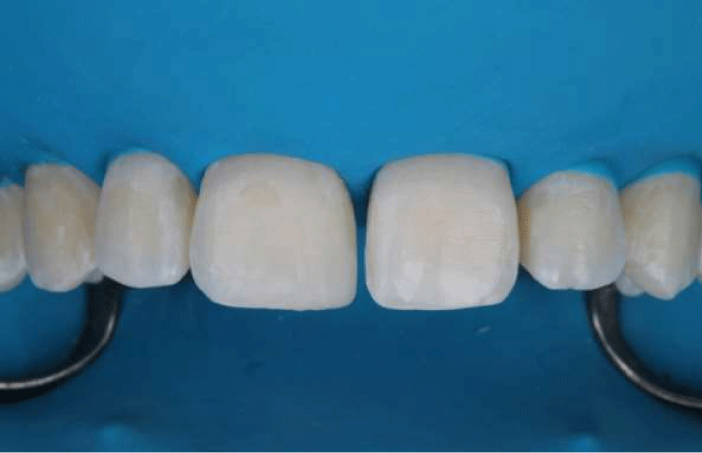
Figure 8: First teeth (11) restored.
After the diastema was closed (Figure 9), the restorations were flattened with abrasive discs (Soflex, 3M ESPE, St. Paul, MN, USA) and polished with silicone points (Enhance, Dentsply Sirona, Charlotte, NC, EUA) followed by the felt disc with a diamond polishing paste (Diamond Excel, FGM, Joinville, SC, Brazil) (Figure 10).
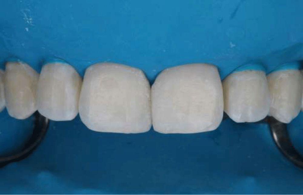
Figure 9: The view of diastema closed before polishing.
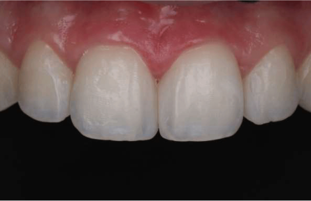
Figure 10: The view of diastema closed after polishing.
At the follow-up three years later, no discolorations or fractures were detected on the teeth and restorations and the patient was completely satisfied (Figures 11 and 12). Despite the color stability was maintained after 3-year follow-up, the proservation indicated the need for anatomy repair and repolishing of the restorations.
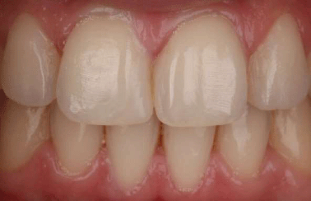
Figure 11: The view of restoration at 3-year recall.
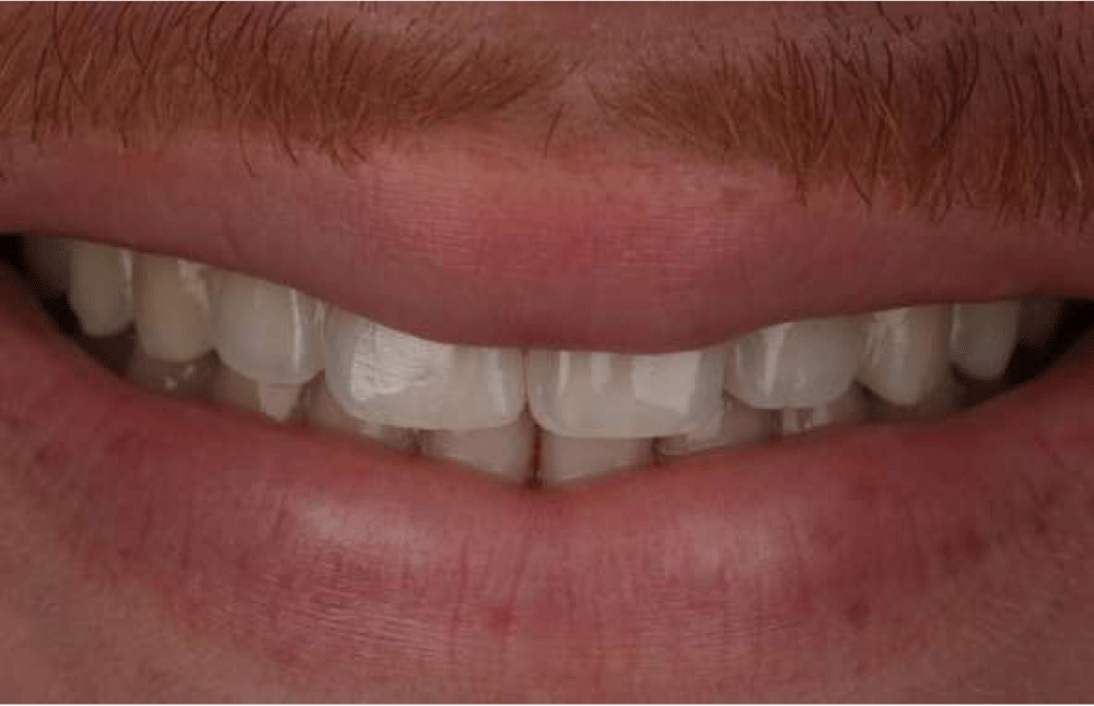
Figure 12: The view of the smile at 3-year recall.
With the aim to reduce tooth sensitivity while maintaining whitening efficacy, the incorporation of a titanium dioxide doped with nitrogen (TIO_N) photo-catalyst into lower concentrations of HP (3.5% – 6%) was proposed [21,23]. In the present case report, the 6% HP gel with TIO_N associated with hybrid light (violet LED/Diode Laser) successfully whitened teeth (from A2 to A1 of the VITA classical shade scale) and the patient did not report any sensitivity during or after the treatment.
Studies [24,25] reported that the use of a violet LED light system associated with low-concentrated bleaching agents is a good option to fulfill the rising demand for aesthetics, promoting efficient tooth bleaching without side effects during and post-treatment, suggesting that the use of low-concentrated HP bleaching agents should be the first choice for in-office bleaching [14,26]. Fernandez, et al. [19] (2017) and Vildosola, et al. [20] (2017) reported that the 6% HP bleaching was effective and remained effective at nine months and 1-year recall, respectively. These studies also reported that patients were satisfied with the bleaching procedure and it had a positive impact on esthetic perception and a positive psychosocial impact at the nine-month recall. The result of the present report showed that the bleaching efficacy was stable at a 3-year recall.
For the diastema closure, as it has many different etiologies such as genetics factors, hypertrophic labial frenum and its insertion, oral habits, anterior traumatic bite and abnormalities in the number or size of teeth [27], an accurate diagnosis of the etiology of the maxillary midline diastema is the key to an effective treatment [28]. In this case, the patient presented a high labial frenal attachment and a labial frenectomy was done before the restoration treatment.
The direct resin composite restoration was chosen to close the diastema between 11 and 21 teeth because the patient was young and this procedure is considered effective, conservative, and predictive and there is no need for cavity preparation [1,29]. In addition, it allows the patient to obtain the result at only one appointment, requiring a shorter treatment time and a relatively lower cost. Another advantage of resin composite restorations is the possibility of repairing small failures [29,30]. Partial restoration durability, color matching with the natural tooth color and marginal adaptation are important points to consider at follow-up [31,32]. In the present study, the restoration remained stable over time and the patient was satisfied at the 3-year follow-up.
Bleaching with a low-concentrated HP gel (6% H2O2) associated with hybrid light (violet LED/Diode Laser) was effective and the result remained stable over 3 years. Also, the resin composite used for diastema closure was stable over time, the shine was obtained by polishing and the color was maintained. At the 3-year follow-up, there were no differences between the resin and teeth and no discolorations or fractures were detected. The patient was completely satisfied. So, the bleaching protocol using low concentrations of HP associated with hybrid light is an interesting option for a young patient, once it is conservative, safe and presents longevity over time.
Ethical considerations
The present study was approved by the Ethics Committee in Research of the Bauru School of Dentistry, University of São Paulo (process n. 68666822.0.0000.5417) and the patient signed the free and informed consent form.
- Hwang SK, Ha JH, Jin MU, Kim SK, Kim YK. Diastema closure using direct bonding restorations combined with orthodontic treatment: a case report. Restor Dent Endod. 2012 Aug;37(3):165-9. doi: 10.5395/rde.2012.37.3.165. Epub 2012 Aug 29. PMID: 23429455; PMCID: PMC3569402.
- Borges AB, Zanatta RF, Barros AC, Silva LC, Pucci CR, Torres CR. Effect of hydrogen peroxide concentration on enamel color and microhardness. Oper Dent. 2015 Jan-Feb;40(1):96-101. doi: 10.2341/13-371-L. Epub 2014 Aug 19. PMID: 25136902.
- Gallinari MO, Fagundes TC, da Silva LM, de Almeida Souza MB, Barboza A, Briso A. A New Approach for Dental Bleaching Using Violet Light With or Without the Use of Whitening Gel: Study of Bleaching Effectiveness. Oper Dent. 2019 Sep/Oct;44(5):521-529. doi: 10.2341/17-257-L. Epub 2019 Apr 25. PMID: 31021692.
- Mondelli R, Rizzante F, Rosa ER, Borges A, Furuse AY, Bombonatti J. Effectiveness of LED/Laser Irradiation on In-Office Dental Bleaching after Three Years. Oper Dent. 2018 Jan/Feb;43(1):31-37. doi: 10.2341/16-208-C. PMID: 29284097.
- Eimar H, Siciliano R, Abdallah MN, Nader SA, Amin WM, Martinez PP, Celemin A, Cerruti M, Tamimi F. Hydrogen peroxide whitens teeth by oxidizing the organic structure. J Dent. 2012 Dec;40 Suppl 2:e25-33. doi: 10.1016/j.jdent.2012.08.008. Epub 2012 Aug 24. PMID: 22925924.
- Gallinari MO, Cintra LTA, Souza MBA, Barboza ACS, Esteves LMB, Fagundes TC, Briso ALF. Clinical analysis of color change and tooth sensitivity to violet LED during bleaching treatment: A case series with split-mouth design. Photodiagnosis Photodyn Ther. 2019 Sep;27:59-65. doi: 10.1016/j.pdpdt.2019.05.016. Epub 2019 May 20. PMID: 31121330.
- Panhoca VH, de Oliveira BP, Rastelli ANS, Bagnato VS. Dental Bleaching Using Violet Light Alone: Clinical Case Report. Dentistry. 2017; 7(11):459. doi: 10.4172/2162-1122.1000459.
- Bersezio C, Estay J, Jorquera G, Peña M, Araya C, Angel P, Fernández E. Effectiveness of Dental Bleaching With 37.5% and 6% Hydrogen Peroxide and Its Effect on Quality of Life. Oper Dent. 2019 Mar/Apr;44(2):146-155. doi: 10.2341/17-229-C. Epub 2018 Oct 10. PMID: 30517065.
- Kutuk ZB, Ergin E, Cakir FY, Gurgan S. Effects of in-office bleaching agent combined with different desensitizing agents on enamel. J Appl Oral Sci. 2018 Nov 8;27:e20180233. doi: 10.1590/1678-7757-2018-0233. PMID: 30427477; PMCID: PMC6223786.
- Faraoni-Romano JJ, Da Silveira AG, Turssi CP, Serra MC. Bleaching agents with varying concentrations of carbamide and/or hydrogen peroxides: effect on dental microhardness and roughness. J Esthet Restor Dent. 2008;20(6):395-402; discussion 403-4. doi: 10.1111/j.1708-8240.2008.00216.x. PMID: 19120787.
- de Almeida LC, Soares DG, Gallinari MO, de Souza Costa CA, Dos Santos PH, Briso AL. Color alteration, hydrogen peroxide diffusion, and cytotoxicity caused by in-office bleaching protocols. Clin Oral Investig. 2015 Apr;19(3):673-80. doi: 10.1007/s00784-014-1285-3. Epub 2014 Jul 19. PMID: 25035067.
- Klaric E, Rakic M, Sever I, Milat O, Par M, Tarle Z. Enamel and Dentin Microhardness and Chemical Composition After Experimental Light-activated Bleaching. Oper Dent. 2015 Jul-Aug;40(4):E132-41. doi: 10.2341/14-148-L. Epub 2015 Mar 6. PMID: 25748206.
- Mondelli RFL, Gabriel TRCG, Rizzante FAP, Magalhães AC, Bombonatti JFS, Ishikiriama SK. Do different bleaching protocols affect the enamel microhardness? Eur J Dent. 2015 Jan-Mar;9(1):25-30. doi: 10.4103/1305-7456.149634. PMID: 25713480; PMCID: PMC4319295.
- Grazioli G, Valente LL, Isolan CP, Pinheiro HA, Duarte CG, Münchow EA. Bleaching and enamel surface interactions resulting from the use of highly-concentrated bleaching gels. Arch Oral Biol. 2018 Mar;87:157-162. doi: 10.1016/j.archoralbio.2017.12.026. Epub 2017 Dec 28. PMID: 29304422.
- Lago ADN, Ferreira WDR, Furtado GS. Dental bleaching with the use of violet light only: Reality or Future? Photodiagnosis Photodyn Ther. 2017 Mar;17:124-126. doi: 10.1016/j.pdpdt.2016.11.014. Epub 2016 Nov 27. PMID: 27902926.
- GDC, Position Statement on Tooth Whitening, in: GD Council (Ed.) https://www.gdc-uk.org/docs/default-source/what-is-the-legal-position/tooth-whitening-position-statementf08f3f9796b446d88663d00f035d4c13.pdf?sfvrsn=16f71e9_7, 2016.
- Alkahtani R, Stone S, German M, Waterhouse P. A review on dental whitening. J Dent. 2020 Sep;100:103423. doi: 10.1016/j.jdent.2020.103423. Epub 2020 Jun 29. PMID: 32615235.
- Kury M, Wada EE, Silva DPD, Tabchoury CPM, Giannini M, Cavalli V. Effect of violet LED light on in-office bleaching protocols: a randomized controlled clinical trial. J Appl Oral Sci. 2020;28:e20190720. doi: 10.1590/1678-7757-2019-0720. Epub 2020 May 18. PMID: 32428059; PMCID: PMC7213781.
- Fernández E, Bersezio C, Bottner J, Avalos F, Godoy I, Inda D, Vildósola P, Saad J, Oliveira OB Jr, Martín J. Longevity, Esthetic Perception, and Psychosocial Impact of Teeth Bleaching by Low (6%) Hydrogen Peroxide Concentration for In-office Treatment: A Randomized Clinical Trial. Oper Dent. 2017 Jan/Feb;42(1):41-52. doi: 10.2341/15-335-C. Epub 2016 Aug 29. PMID: 27571237.
- Vildósola P, Vera F, Ramírez J, Rencoret J, Pretel H, Oliveira OB Jr, Tonetto M, Martín J, Fernández E. Comparison of Effectiveness and Sensitivity Using Two In-Office Bleaching Protocols for a 6% Hydrogen Peroxide Gel in a Randomized Clinical Trial. Oper Dent. 2017 May/Jun;42(3):244-252. doi: 10.2341/16-043-C. PMID: 28467262.
- Martín J, Vildósola P, Bersezio C, Herrera A, Bortolatto J, Saad JR, Oliveira OB Jr, Fernández E. Effectiveness of 6% hydrogen peroxide concentration for tooth bleaching—A double-blind, randomized clinical trial. J Dent. 2015 Aug;43(8):965-72. doi: 10.1016/j.jdent.2015.05.011. Epub 2015 Jun 6. PMID: 26057085.
- Rastelli ANS, Dias HB, Carrera ET, de Barros ACP, Dos Santos DDL, Panhóca VH, Bagnato VS. Violet LED with low concentration carbamide peroxide for dental bleaching: A case report. Photodiagnosis Photodyn Ther. 2018 Sep;23:270-272. doi: 10.1016/j.pdpdt.2018.06.021. Epub 2018 Jun 28. PMID: 29964222.
- Moncada G, Sepúlveda D, Elphick K, Contente M, Estay J, Bahamondes V, Fernandez E, Oliveira OB, Martin J. Effects of light activation, agent concentration, and tooth thickness on dental sensitivity after bleaching. Oper Dent. 2013 Sep-Oct;38(5):467-76. doi: 10.2341/12-335-C. Epub 2013 Feb 7. PMID: 23391030.
- Brugnera AP, Nammour S, Rodrigues JA, Mayer-Santos E, de Freitas PM, Brugnera A Junior, Zanin F. Clinical Evaluation of In-Office Dental Bleaching Using a Violet Light-Emitted Diode. Photobiomodul Photomed Laser Surg. 2020 Feb;38(2):98-104. doi: 10.1089/photob.2018.4567. Epub 2019 Aug 22. PMID: 31436475.
- Bortolatto JF, Trevisan TC, Bernardi PS, Fernandez E, Dovigo LN, Loguercio AD, Batista de Oliveira Junior O, Pretel H. A novel approach for in-office tooth bleaching with 6% H2O2/TiO_N and LED/laser system-a controlled, triple-blinded, randomized clinical trial. Lasers Med Sci. 2016 Apr;31(3):437-44. doi: 10.1007/s10103-016-1866-2. Epub 2016 Jan 21. PMID: 26796706.
- Soares DG, Ribeiro AP, da Silveira Vargas F, Hebling J, de Souza Costa CA. Efficacy and cytotoxicity of a bleaching gel after short application times on dental enamel. Clin Oral Investig. 2013 Nov;17(8):1901-9. doi: 10.1007/s00784-012-0883-1. Epub 2012 Dec 6. PMID: 23224040.
- Oesterle LJ, Shellhart WC. Maxillary midline diastemas: a look at the causes. J Am Dent Assoc. 1999 Jan;130(1):85-94. doi: 10.14219/jada.archive.1999.0033. PMID: 9919036.
- Abraham R, Kamath G. Midline diastema and its aetiology--a review. Dent Update. 2014 Jun;41(5):457-60, 462-4. doi: 10.12968/denu.2014.41.5.457. PMID: 25073229.
- Demirci M, Tuncer S, Öztaş E, Tekçe N, Uysal Ö. A 4-year clinical evaluation of direct composite build-ups for space closure after orthodontic treatment. Clin Oral Investig. 2015 Dec;19(9):2187-99. doi: 10.1007/s00784-015-1458-8. Epub 2015 Mar 24. PMID: 25802222.
- Wolff D, Kraus T, Schach C, Pritsch M, Mente J, Staehle HJ, Ding P. Recontouring teeth and closing diastemas with direct composite buildups: a clinical evaluation of survival and quality parameters. J Dent. 2010 Dec;38(12):1001-9. doi: 10.1016/j.jdent.2010.08.017. Epub 2010 Sep 6. PMID: 20826192.
- Pereira Sanchez N, Powers JM, Paravina RD. Instrumental and visual evaluation of the color adjustment potential of resin composites. J Esthet Restor Dent. 2019 Sep;31(5):465-470. doi: 10.1111/jerd.12488. Epub 2019 May 16. PMID: 31095870.
- Korkut B, Türkmen C. Longevity of direct diastema closure and recontouring restorations with resin composites in maxillary anterior teeth: A 4-year clinical evaluation. J Esthet Restor Dent. 2021 Jun;33(4):590-604. doi: 10.1111/jerd.12697. Epub 2020 Dec 23. PMID: 33354867.