
More Information
Submitted: Jujy 25, 2026 | Accepted: August 01, 2026 | Published: August 03, 2026
Citation: Hugues JCO. Dental Microscope-Assisted and Indirect Ultrasonic Technique for Retrieval of Separated Endodontic Instruments: A Case Report. J Clin Adv Dent. 2026; 10(1): 7-14. Available from:
https://dx.doi.org/10.29328/journal.jcad.1001054
DOI: 10.29328/journal.jcad.1001054
Copyright license: © 2026 Hugues JCO. This is an open access article distributed under the Creative Commons Attribution License, which permits unrestricted use, distribution, and reproduction in any medium, provided the original work is properly cited.
Keywords: File retrieval; Indirect Uultrasonic technique; Dental microscope; Magnification; Complex endodontics

Dental Microscope-Assisted and Indirect Ultrasonic Technique for Retrieval of Separated Endodontic Instruments: A Case Report
Juan Carlos Ortiz Hugues*
AMED President, Dental Microscope Training Director of Endodontics, USA
*Corresponding author: Juan Carlos Ortiz Hugues, AMED President, Dental Microscope Training Director of Endodontics, USA, Email: [email protected]
Endodontic procedures pose significant challenges during both the diagnostic and treatment phases of root canal therapy (RCT). Instrument separation or fracture is a potential complication during canal shaping, especially when utilizing rotary nickel-titanium (NiTi) files.
The dental microscope is a required magnification tool in United States endodontic training programs and is considered an essential component of the endodontist’s clinical toolkit. The improved visual information and resolution provided by the microscope’s lighting and adjustable magnification allow clinicians to address complex challenges in deep, narrow canal regions and to manage obstructions that may compromise the success of root canal treatment.
The decision to bypass or retrieve a separated instrument should be informed by established clinical principles. The selection of appropriate tools and techniques for file retrieval must be consistent with these principles to ensure precise and effective treatment.
This case report outlines a systematic approach to file retrieval during complex root canal treatment, guided by established clinical principles, continuous magnification, and the use of Sonic Steel probes designed for the indirect ultrasonic file retrieval technique. The clinical effectiveness of the indirect ultrasonic technique, in combination with ultrasound, is demonstrated in this case report.
Endodontic procedures present several potential complications, with instrument fracture representing a significant concern [1]. The literature reports an incidence rate of 0.25% to 6% for instrument fractures [2]. Nickel-titanium files demonstrate a fracture rate of 1.3% to 10% [3,4]. which exceeds that of stainless steel manual files. Fractures most frequently occur in curved molar canals and narrow, constricted canals due to torsional stress or instrument fatigue [51].
Although instrument fracture is a relatively common procedural complication, it accounts for less than 1% of all endodontic treatment failures. Despite its low incidence, most practitioners are likely to encounter this complication during their careers. Instrument fracture is recognized as a significant source of stress among endodontists [6].
These failures are frequently attributed to the following factors: [7]
- Complex root canal anatomy, including canal curvature, narrow canals, and calcified canals
- Operator error, such as improper technique, diagnostic errors, inappropriate instrument selection, excessive or forceful use of instruments, inadequate access, and suboptimal file properties.
In these scenarios, practitioners must be prepared to manage the incident by considering two primary clinical factors. First, establishing a proper glide path with manual or appropriately selected rotary or oscillatory instruments [8], tailored to the specific complexities of canal anatomy such as curves, bicurvatures, or narrowness, increases the likelihood of bypassing the separated instrument or preventing flute engagement with the canal wall dentin [9]. This approach facilitates fragment retrieval, as the elliptical shape of most canals provides additional space for bypassing or retrieval with less resistance.
Second, file retrieval should not be attempted without a dental microscope, as effective treatment is limited to what is visible. In these situations, clinicians should prioritize visualization to enhance the likelihood of successful fragment retrieval [10].
Postgraduate endodontic programs in the United States require the use of microscopes for specialty training. Ergonomic use of magnification tools is essential for both comfort and efficiency during root canal procedures. The optimized coaxial lighting and multiple magnification levels provided by dental microscopes offer clinicians the visual information and resolution necessary to operate in dark, narrow spaces deep within the canals [11].
During file retrieval, detection of the fragment is aided by the metal’s brightness and its position. The use of specially designed ultrasonic tips, probes, tubes, or loops can facilitate fragment retrieval when used in accordance with established safety guidelines [12].
Continuous microscope-assisted magnification is essential for obtaining accurate visual information during file retrieval, regardless of the technique employed. The primary objective is to maintain a clear visualization of the fragment to facilitate its stepwise removal. This is achieved by applying EDTA, alcohol, or hypochlorite to keep the canal moist, remove the smear layer, and clean the area, and by cooling the tooth structure during ultrasonic vibration, which is commonly used for this procedure [13].
Nickel-titanium alloys have represented a significant advancement in endodontics, enabling more precise and efficient shaping protocols due to their flexibility. However, these alloys are also a primary source of procedural accidents, particularly file breakage during root canal treatment. The most significant technical causes of file breakage are cyclic fatigue and torsional stress. Cyclic fatigue results from repeated bending and unbending stresses on the instrument surface, especially in curved or narrowed canals. Torsional stress arises from twisting forces, particularly when instruments are subjected to apical pressure or remain in continuous rotation within curved or narrow canals [14].
Additional sources of file breakage include manufacturing defects, which can cause instruments to fracture under minimal stress during use. Failure to discard instruments after use also increases the risk of breakage [15]. Both factors contribute to procedural accidents, underscoring the need for practitioners to strictly adhere to established protocols and principles when using nickel-titanium files to minimize such incidents.
The initial radiograph should be analyzed to assess the complexity of the root canal treatment. Key factors include root curvature, presence of multiple curvatures, tooth position, patient limitations in mouth opening, and the degree of canal calcification. This evaluation informs the selection of an appropriate shaping protocol, instrument choice, and sequence. It enables the clinician to determine the optimal maximum taper and select files with suitable thermal, mechanical, or stiffness properties, thereby optimizing the procedure and minimizing the risk of instrument fracture [16].
Addressing the cervical third of the root is essential. Pre-flaring the dentin triangle and establishing a straight path with size 10 files from the cervical to the apical third reduces stress on subsequent instruments and facilitates reaching working lengths [17]. Incorporating glide path preparation, irrigation activation, and instrument recapitulation from the outset enhances the efficiency of cleaning and shaping. This approach also increases the likelihood of successfully bypassing a separated instrument within a previously cleaned and shaped canal [18].
In the event of file breakage during root canal treatment, the practitioner must follow a structured decision-making process. The initial step is to inform the patient of the incident, as this is a recognized risk associated with endodontic procedures. Practitioners should assess canal anatomy through initial radiographic diagnosis and by negotiating the canal with a #10 K-File. The chosen technique should then correspond to the complexity encountered during the shaping stage of root canal therapy [19].
File breakage may occur in the cervical, middle, or apical third of the canal, or after a canal curvature. Additional complicating factors include long, narrow canals and chronic infection. These scenarios require the endodontist to carefully determine the most appropriate approach and technique for file retrieval or bypass [20].
The preferred initial strategy is to attempt to bypass the fractured instrument, particularly if prior canal preparation has established a glide path [21]. In such cases, the instrument may be less firmly seated in the canal, allowing the use of a #8 or #10 K-File to bypass the fragment and access the apical region for effective cleaning and shaping. Leaving the fragment as part of the root canal filling reduces the risk of excessive dentin removal or perforation, offering both biological and structural advantages over the long term [22].
If bypassing the file is not feasible, retrieval should be considered according to established criteria, including the fragment’s location, root length, instrument type, and taper. The likelihood of successful retrieval varies depending on these factors [23].
Fragment location in the root: If the file is located beyond the curve, the chances of successful retrieval are lowest, with a high risk of perforation or damage to the root structure [24].
Instrument Taper: An increase in taper, file caliber, shape, flute design, or helix angle results in greater engagement with the inner dentinal walls, thereby increasing procedural complexity and risk [25].
Length of the root: If the fragment is located in the middle or apical third of a long root, retrieval requires the use of longer, thinner, and more delicate instruments. This process necessitates high magnification, advanced motor skills, and precise force application to dislodge the file from the canal walls during retrieval [26].
A variety of specialized instruments and techniques for file retrieval are commercially available. These methods may be combined to improve the overall effectiveness of the retrieval process [27].
After achieving clear visualization of the fragment using appropriate magnification, accurate localization facilitates direct management of the fragment and reduces associated risks. The next step involves creating a platform at the coronal aspect of the fragment and exposing 2–3 mm of its top surface, which enables the application of subsequent techniques or their combination [28].
Direct Ultrasonic Technique
Ultrasonic units equipped with specially designed, long, thin tips for instrument retrieval can create space between the fragment and the dentin walls. These tips facilitate bypassing or releasing the instrument, typically by applying a counterclockwise motion [29].
Indirect Ultrasonic Technique
Specially designed sonic steel alloy probes with diamond-coated thin tips are commercially available. When activated by an ultrasonic tip, these probes generate secondary, less intense vibrations, resulting in less aggressive dentin removal. The probe tip’s flexibility allows it to bend toward the fractured instrument, providing versatility in creating space for bypass or retrieval with reduced risk [30](Figure 1).
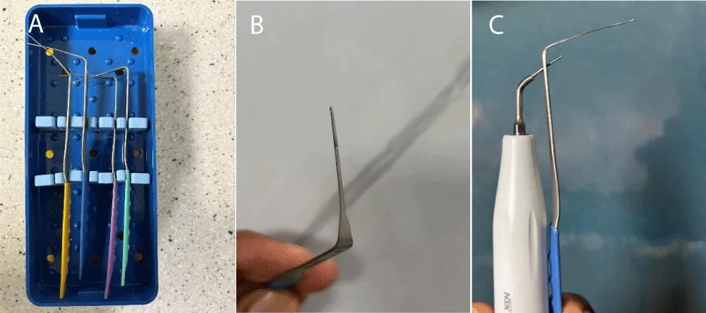
Figure 1: Sonic Steel diamond-coated probes, Laschal FXP System. Laschal Surgical Inc (A) Kit with various angulations. (B) Diamond-coated tip. (C) An ultrasonic tip is inserted into the probe to transmit indirect ultrasonic energy.
Tube Technique
These instruments consist of a hollow tube and a specialized extractor designed to grip and remove the fragment. Multiple calibers are available to accommodate varying canal widths [31].
Lasso Technique
This instrument incorporates a specially designed loop that secures the fragment and facilitates its extraction [32].
File Removal Kits
Commercially available kits provide comprehensive instrument systems, including trephines for platform creation, ultrasonic tips, loops, tweezers, and tube systems of various calibers [33].
Selection of technique and instrumentation is influenced by cost, operator preference, and adaptability. In some cases, multiple techniques may be utilized concurrently.
A 56-year-old female patient presented with acute, spontaneous symptoms in tooth number 18 that intensified upon biting. Clinical examination identified a leaking crown and an aged composite restoration on the occlusal surface.
Periapical radiography demonstrated an endodontic file retained in the middle to apical third of the mesial root, at the onset of the curvature. Figure 2.1 The previous clinician neither documented nor explained the retained file and did not complete the initiated root canal treatment. A small periapical radiolucency was also observed in the distal root.
The presumptive diagnosis was chronic apical periodontitis. The proposed treatment plan included root canal therapy (RCT), core build-up, and placement of a new crown.
During the procedure, a dental microscope and multiple monitors enabled real-time observation of the treatment, allowing the clinician to provide detailed, step-by-step explanations.
The initial steps involved removal of the crown, placement of a rubber dam, and elimination of the compromised composite restoration and underlying carious tissue. Figure 2,3 Contamination within the pulp chamber was observed following removal. The DTE Ultrasonic Scaler, configured for endodontic procedures with E5 Helse and E3D Helse ultrasonic tips at medium power (5), was employed to eliminate residual composite from the chamber floor and walls. Sodium hypochlorite (NaClO), activated with the E5 ultrasonic tip, was utilized to irrigate the chamber until complete decontamination was achieved.
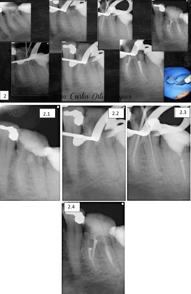
Figure 2: Sequential periapical radiographs illustrating file retrieval and management of root canal treatment (RCT). Figures 2.1, 2.2, and 2.3 Present individual periapical radiographs: 2.1 shows the initial radiograph, 2.2 displays the retrieved instrument, and 2.3 depicts the final endodontic obturation radiograph.
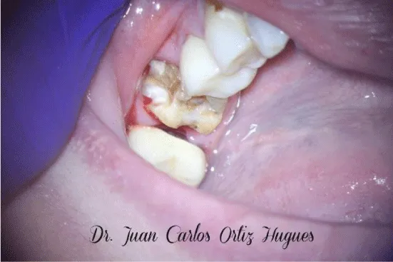
Figure 3: Disassembled crown and composite removal.
The mesiobuccal (MB), mesiolingual (ML), and distal (D) canals were located using a DG-15 explorer. Initial scouting of the canals and determination of the glide path were performed with size 08 and 10 files, with the chamber flooded with NaClO. Pre-flaring of the cervical triangle of the roots was accomplished using size 10, 15, and 20 files in the cervical third, along with an orifice shaper, to remove the triangle and optimize working length instrumentation. The working length was determined after preparation of the cervical third of the root, using a K-File #10 in the mesiolingual canal and a K-File #15 in the distal canal. The measurement was verified with an apex locator (Pixi, Eighteeth, Changzhou Sifary Medical Technology Co) and periapical radiography.
After confirming the working length radiographically, cleaning and shaping were initiated in the ML and D canals, with the MB canal reserved for the fragmented instrument last. The ML and D canals were initially instrumented manually at working length with size 15, 20, and 25 K-files, with size 10 files used for recapitulation to maintain the glide path. NaClO irrigation with passive ultrasonic irrigation (PUI) activation was performed between each file, using a size 10 file activated with the E5 ultrasonic tip to create cavitation within the canal. Following manual instrumentation, a rotary file sequence (15.04, 20.05, 25.04, 30.04) was employed, with size 10 file recapitulation and 17% EDTA used with each rotary instrument.
After cleaning and shaping the ML and D canals, the instrument retrieval protocol was initiated. The cervical third of the MB root canal was managed with NaClO PUI activation, followed by 17% EDTA PUI activation and isopropyl alcohol to dry the canal. High magnification with the dental microscope was used to clearly identify the coronal portion of the fragmented instrument. Continuous magnification inspection was maintained throughout the retrieval process. Figure 4. Once the coronal portion was visualized, a Gates-Glidden #1 was modified by cutting at the equator of the active part to create a flat platform, which was then carefully introduced to the depth of the canal where the fragment was located. This platform enlarged the orifice, providing visual and dimensional space for the Sonic Steel diamond-coated flexible probe to work around the instrument incrementally.
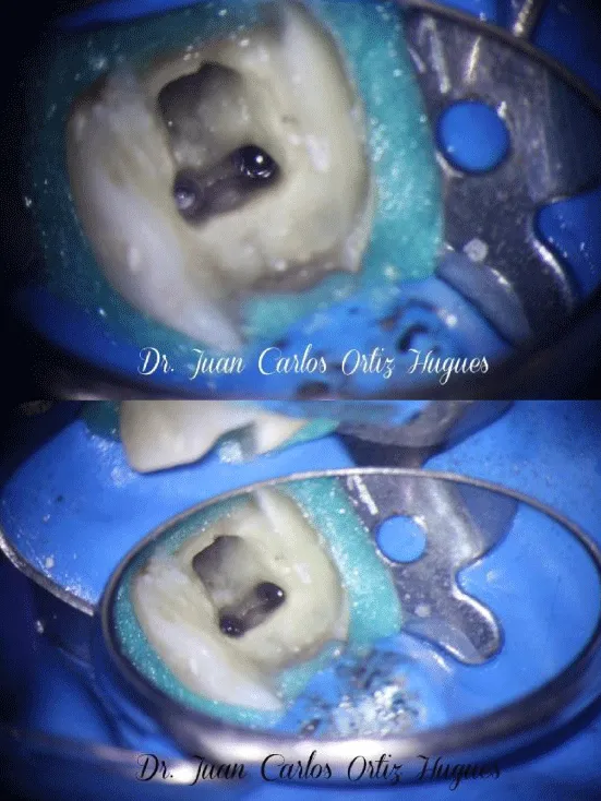
Figure 4: High magnification using a dental microscope enabled clear identification of the coronal portion of the fragmented instrument. Magnification was continuously maintained throughout the retrieval process.
The Sonic Steel Probe Instrument (FXP; Laschal Surgical, New York, USA) comprises four instruments, each with a distinct angulation and identified by blue, yellow, purple, or green colors. The blue and yellow instruments were selected according to the angulation required for the mesiobuccal (MB) root of the specific tooth. The FXP instrument contains an orifice at its top designed for placement of the E5 Ultrasonic Tip. Figure 5. Upon activation, the tip transmits energy to the FXP probe, generating a secondary, more controlled force and thereby reducing aggressive canal wear. The probe’s alloy tip was bent and oriented toward the coronal portion of the instrument, which enabled precise positioning between the inner canal wall and the instrument in a counterclockwise direction. Additional procedures included passive ultrasonic irrigation (PUI), alcohol, and paper points, with continuous inspection under magnification. The fragment was lodged against the dentinal walls at the beginning of the curvature, with approximately 6 mm of the instrument engaged, which made retrieval challenging.
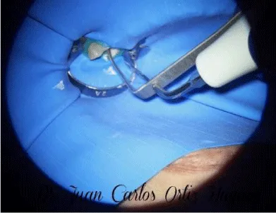
Figure 5: Indirect ultrasonic technique for file retrieval using a Sonic Steel diamond-coated flexible probe activated by an ultrasonic E5 tip.
Supplementary Video: View the dental microscope-assisted indirect ultrasonic file retrieval video. https://youtu.be/47b43um3d-g?si=u3GikZHBurkM5UG8
Given the complexity of instrument retrieval and the patient’s restricted mouth opening and discomfort, calcium hydroxide (Ultracal, Ultradent) was placed in all three canals for one week, followed by the application of temporary cement. Instrument retrieval was scheduled for a subsequent appointment one week later.
At the second appointment, the patient’s pain and discomfort had decreased to minimal levels. We proceeded with the indirect ultrasonic technique, successfully loosening the fragment by repeating the established protocol. The file was then captured in the pulp chamber using Stieglitz forceps. Subsequently, the canal was glide-pathed with a size 10 K-file. The working length was determined using an apex locator and confirmed by periapical X-ray. Continuous activation of sodium hypochlorite (NaClO) was performed using passive ultrasonic irrigation (PUI), and the same rotary file protocol was employed as for the mesiolingual (ML) and distal (D) canals (Figure 6).
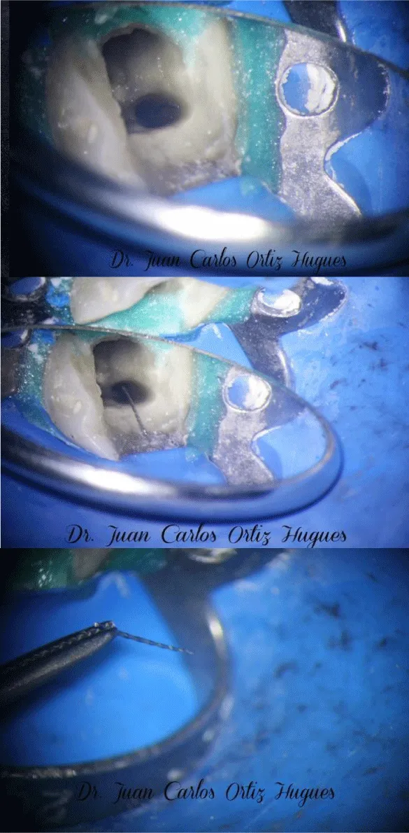
Figure 6: Successful File Retrieval.
After drying all three canals with isopropyl alcohol and paper points, we employed the Hybrid Tagger Obturation technique using Sealapex cement. Gutta-percha was thermomechanically condensed with a Guttacondensor #35 instrument. Teflon and Cavit materials were placed on the coronal aspect of the tooth, preparing it for build-up by the referring dentist (Figures 2.3, 2.4, 7).
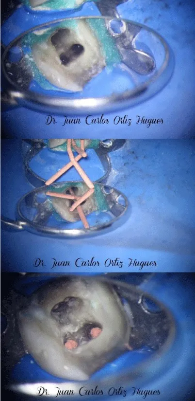
Figure 7: Distal root following removal of the file, demonstrating final root canal treatment (RCT) filling.
Retrieval of fractured endodontic instruments remains one of the most technically demanding procedures in endodontics. The prognosis of instrument retrieval depends on several factors, including the location of the fractured fragment, canal curvature, root anatomy, remaining dentin thickness, instrument design, and the clinician’s experience. Instrument separation is a recognized procedural complication, particularly when nickel-titanium (NiTi) rotary instruments are used in complex root canal systems, where cyclic fatigue and torsional stress significantly contribute to fracture occurrence. Previous clinical studies have reported varying incidences of instrument separation, emphasizing the importance of preventive strategies such as appropriate case assessment, glide path preparation, controlled instrumentation, and operator experience [1-9].
Current clinical guidelines recommend that management of fractured instruments should be based on a systematic decision-making process considering the position of the fragment, pre-existing infection, canal accessibility, and the potential risks associated with dentin removal [2,12,19,20].Depending on these clinical factors, the clinician may choose to bypass the fragment, retrieve it, or incorporate it into the final obturation when retrieval presents an unacceptable risk to tooth integrity [21-24].
The present case demonstrates the successful application of the indirect ultrasonic retrieval technique under continuous dental microscope magnification. Magnification and coaxial illumination considerably improve visualization of the operative field, facilitating conservative dentin removal while minimizing the risk of perforation or excessive weakening of the root structure. The routine use of the operating microscope has become an essential component of contemporary endodontic practice and significantly improves the predictability of complex procedures such as fractured instrument retrieval [10,11,13].
Compared with conventional direct ultrasonic techniques, the indirect ultrasonic approach using the Sonic Steel diamond-coated flexible probe provides controlled transmission of ultrasonic energy to the fractured instrument. This indirect vibration reduces aggressive dentin cutting while allowing gradual loosening of the fragment from the canal wall. Similar advantages of ultrasonic-assisted retrieval have been described in previous reports, which demonstrated high clinical success while preserving radicular dentin when appropriate magnification and careful instrumentation were employed [29,30].
Alternative retrieval methods, including tube systems, lasso techniques, and commercially available retrieval kits, have demonstrated satisfactory clinical outcomes in selected cases. However, each technique possesses inherent advantages and limitations that depend largely on fragment location, canal anatomy, and operator proficiency. Tube techniques are generally more effective when sufficient coronal exposure of the fractured fragment is available, whereas lasso systems may be useful in relatively straight canals but are often difficult to manipulate in severely curved canals. Commercial retrieval kits provide comprehensive instrument systems but frequently require greater dentin removal to establish adequate access [23,27,31,33]. Consequently, no single retrieval technique can be regarded as universally superior, and clinicians should individualize treatment according to each clinical situation.
In the present case, the fractured instrument was located at the beginning of the canal curvature, a region generally considered favorable for conservative retrieval when adequate visualization and specialized instruments are available. The use of cervical pre-flaring, glide path preparation, passive ultrasonic irrigation, controlled working-length determination, and staged treatment with calcium hydroxide dressing contributed to safe retrieval while preserving tooth structure. Previous investigations have demonstrated that these procedural strategies significantly improve clinical outcomes while reducing the likelihood of secondary procedural errors [8,17,18,25,26].
The patient’s symptoms resolved after treatment, and successful completion of root canal therapy was achieved without procedural complications. Although this report demonstrates the clinical feasibility of the indirect ultrasonic technique, it represents a single clinical case. Future prospective clinical studies involving larger patient populations and long-term follow-up are required to compare indirect ultrasonic retrieval with other established retrieval techniques regarding success rate, dentin preservation, treatment time, and long-term prognosis [23,27].
Instrument separation represents a recognized risk in endodontic procedures. Employing a shaping protocol informed by anatomical considerations and relevant clinical factors supports effective decision-making about optimal file-retrieval techniques.
No single retrieval technique demonstrates universal superiority. The selection of instruments and methods depends on the clinician’s adaptability and proficiency. In certain cases, combining techniques may yield synergistic outcomes.
The use of the FXP sonic steel probe, in conjunction with substantial expertise in file retrieval, indicates that the indirect ultrasonic technique is both safe and less aggressive compared to the direct ultrasonic technique for file retrieval or instrument bypassing.
Continuous utilization of the dental microscope during complex endodontic procedures is critical for precise management of challenging root canal treatments. Adhering to visualization protocols enhances procedural accuracy, particularly during the retrieval of fractured instruments from deep or narrow canals. This approach minimizes damage to tooth structure by employing specialized instruments, such as a diamond-coated probe activated with indirect ultrasound energy. Additionally, applying controlled high-power steps to the instrument head facilitates safe dislodgement of fractured instruments from canal walls.
In this case, the success of root canal treatment depended on the effective bypass or retrieval of the separated file located at the initial canal curvature, a process characterized by significant complexity. The combination of the indirect ultrasonic technique and incremental magnification with the dental microscope provided the visual information and resolution required to control file position, selectively remove dentin, and safely vibrate the instrument. This strategy enabled efficient file retrieval, canal cleaning, shaping, and obturation.
“This case report was reviewed and approved by the Director of the Dental Clinic Review Board (Protocol Number: 100). Written informed consent was obtained from the patient for the publication of this case report, including all clinical photographs, radiographic images (X-rays), and anonymized personal data for scientific and educational purposes.”
- Gollo EKF, de Almeida Gomes F, Pilownic KJ, Böttcher DE, Vieira CC, Pappen FG. Fracture Risk of Endodontic Files: Clinical Analysis of Reciproc and X1 Blue After Multiple Uses. Aust Endod J. 2025 Aug 6;51(3):699–705. Available from: https://doi.org/10.1111/aej.70003
- Yan F, Gao Y, Wang X, Fan B, Chen Z, Yu Q, et al. Expert consensus on management of instrument separation in root canal therapy. Int J Oral Sci. 2025 Jun 9;17:46. Available from: https://doi.org/10.1038/s41368-025-00372-w
- Knowles KI, Hammond NB, Biggs SG, Ibarrola JL. Incidence of instrument separation using LightSpeed rotary instruments. J Endod. 2006;32(1):14–6. Available from: https://doi.org/10.1016/j.joen.2005.10.008
- Baumann MA, Roth A. Effect of experience on quality of canal preparation with rotary nickel-titanium files. Oral Surg Oral Med Oral Pathol Oral Radiol. 1999;88(6):714–8. Available from: https://doi.org/10.1016/s1079-2104(99)70015-6
- Alfouzan K, Jamleh A. Fracture of nickel titanium rotary instrument during root canal treatment and re‐treatment: A 5‐year retrospective study. August 2017, International Endodontic Journal 51(2):157-163. Available from: https://doi.org/10.1111/iej.12826
- Rao AS, Surana MA, Shah N, Pawar AM. A nationwide cross-sectional study on endodontic instrument fractures and development of a comprehensive classification system. J Conserv Dent Endod. 2025 Sep 5;28(9):916–924. Available from: https://doi.org/10.4103/jcde.jcde_307_25
- Eskibağlar M, Özata MY, Ocak MS, Öztekin F. Investigation of fracture prevalence of instruments used in root canal treatments at a faculty of dentistry: a prospective study. Restor Dent Endod 2023;48(4):e38. Available from: https://doi.org/10.5395/rde.2023.48.e38
- Berutti E, Negro A, Lendini M, Pasqualini D. Influence of Manual Preflaring and Torque on the Failure Rate of ProTaper Rotary Instruments. J. Endod. 2004;30:228–230. Available from: https://doi.org/10.1097/00004770-200404000-00011
- Vouzara T, Chares E, Lyroudia K. Separated instrument in endodontics: Frequency, treatment and prognosis. Balkan Journal of Dental Medicine 22(3):123-132.: Available from: https://doi.org/10.2478/bjdm-2018-0022
- Ruddle CJ. Micro-endodontic nonsurgical retreatment. Dent Clin North Am 1997; 41:429–454.
- Castellucci A. The use of the operating microscope in endodontics. Endodontic Practice. 2004: 27(2), 18–24.
- Fan Y, Gao Y, Wang X, Fan B, Chen Z, Yu Q, et al. Expert consensus on management of instrument separation in root canal therapy. Int J Oral Sci. 2025 Jun 9;17:46. Available from: https://doi.org/10.1038/s41368-025-00372-w
- Kaul R, Gupta R, Chhabra S, Koull R. Dental Operating Microscope-guided Retrieval of Broken Instrument from a Deciduous Molar Using Ultrasonics. Int J Clin Pediatr Dent. 2022;15. Available from: https://doi.org/10.5005/jp-journals-10005-1892
- Pedullà E, Savio FL, La Rosa GRM, Miccoli G, Bruno E, Rapisarda S, et al. Cyclic fatigue resistance, torsional resistance, and metallurgical characteristics of M3 Rotary and M3 Pro Gold NiTi files. Restor Dent Endod. 2018 Apr 23;43(2):e25. Available from: https://doi.org/10.5395/rde.2018.43.e25
- Khosravi MR, Banafshi Z, Ebrahimi R, Jalali R. Exploring dentists’ experiences of endodontic file fracture during root canal treatment: a phenomenological study. BMC Oral Health. 2025 Jul 1;25:963.Available from: https://doi.org/10.1186/s12903-025-06340-4
- Hartmann RC, Fensterseifer M, Peters OA, de Figueiredo JAP, Gomes MS, Rossi-Fedele G. Methods for measurement of root canal curvature: a systematic and critical review. Int Endod J. 2019 Feb;52(2):169-180. Available from: https://doi.org/10.1111/iej.12996
- Ashwini et al. Addressing the cervical third of the root is essential. Pre-flaring the dentin triangle and establishing a straight path with size 10 files from the cervical to the apical third reduces stress on subsequent instruments and facilitates reaching working lengths. J Contemp Dent Pract. 2012 Jul 1;13(4):554-8.
- Ajina M, Billis G, Chong BS. The Effect of Glide Path Preparation on Root Canal Shaping Procedures and Outcomes. Eur Endod J. 2022 Jun 21;7(2):92–105. Available from: https://doi.org/10.14744/eej.2022.97659
- Solomov M. Broken Instruments – Clinical Decision Making Algorithm. Problem Solving in Endodontics, November 2, 2020. Available from: https://www.aae.org/specialty/category/problem-solving-in-endodontics/
- Chandak M, Sarangi S, Dass A, Khubchandani M, Chandak R. Demystifying Failures Behind Separated Instruments: A Review. Cureus. 2022 Sep 26;14(9):e29588. Available from: https://doi.org/10.7759/cureus.29588
- Parveen S, Hossain M, Uddin FU. Plan International. Management of broken instrument by file bypass technique. February 2017Bangabandhu Sheikh Mujib Medical University Journal 10(1):41-43. Available from: https://doi.org/10.3329/bsmmuj.v10i1.31305
- Adl A, Shahravan A, Farshad M, Honar S. Success Rate and Time for Bypassing the Fractured Segments of Four NiTi Rotary Instruments. Iran Endod J. 2017 Summer;12(3):349–353. Available from: https://doi.org/10.22037/iej.v12i3.16866
- Lakshmaiah D, Kumar JR, Sakthi N, Karunakaran J, Vishwanath S. The Management of Fractured Dental Instruments: A Case Series. Cureus. 2023 Nov 20;15(11):e49132. Available from: https://doi.org/10.7759/cureus.49132
- Nevares G, Cunha RS, Zuolo ML, da Silveira Bueno CE. Success Rates for Removing or Bypassing Fractured Instruments: A Prospective Clinical Study. April 2012. Journal of Endodontics 38(4):442-4. Available from: https://doi.org/10.1016/j.joen.2011.12.009
- Sung HJ, Ha JH, Kim SK. Influence of taper on the screw-in effect of nickel-titanium rotary files in simulated resin root canal. JKACD Volume 35, Number 5, 2010. Available from: https://doi.org/10.5395/JKACD.2010.35.5.380
- Portigliatti R, Lizzi EPC, Rodríguez PA. Predictive factors in the retrieval of endodontic instruments: the relationship between the fragment length and location. Restor Dent Endod. 2024 Sep 9;49(4):e35. Available from: https://doi.org/10.5395/rde.2024.49.e35
- Gopal P, Rao HM, Keshava Prasad BS, Umesh S. Instrument Retrieval in Endodontics: A Literature Review. September 2025. Asian Journal of Dental and Health Sciences 5(3):13-15. Available from: https://doi.org/10.22270/ajdhs.v5i3.133
- Rouggani FZ, Belkamel A. Instrumental fracture in endodontics: a short review," Health Sciences: Vol. 4: Iss. 1, Article 13.2023; Available from: https://doi.org/10.56264/2658-865X.1090
- Chhina H, Hans MK, Chander S. Ultrasonics: A Novel Approach for Retrieval of Separated Instruments. J Clin Diagn Res. 2015 Jan 1;9(1): ZD18–ZD20. Available from: https://doi.org/10.7860/jcdr/2015/11056.5473
- Ortiz Hugues JC. Mastering the art of broken file retrieval in endodontics. Decisions in Dentistry. 2005; 11(3), 32–35.
- Olczak K, Grabarczyk J, Szymański W. Removing fractured endodontic files with a tube technique — the strength of the glued joint: tube-endodontic file setup. Materials. 2023;16:4100. Available from: https://doi.org/10.3390/ma16114100
- Muiño JMS, Fulvi AN, Gimenez M, Rullan JRM. Outside-In SingleeLasso Loop Technique for Meniscal Repair: Fast, Economic, and Reproducible Arthroscopy Techniques, 2018: 7:11. e1191-e1196. Available from: https://doi.org/10.1016/j.eats.2018.08.001
- Pruthi PJ, Nawal RR, Talwar S, Verma M. Comparative evaluation of the effectiveness of ultrasonic tips versus the Terauchi file retrieval kit for the removal of separated endodontic instruments. Restor Dent Endod. 2020 Feb 6;45(2):e14. Available from: https://doi.org/10.5395/rde.2020.45.e14